The Role of Peptide Arrays in Screening Research
Peptide arrays are defined as solid-phase platforms that immobilize thousands of discrete peptide sequences on a single functionalized surface to enable parallel, high-throughput screening against biological targets. The role of peptide arrays in screening spans drug discovery, epitope mapping, biomarker identification, and antibody specificity profiling, making them one of the most productive tools in contemporary biomedical research. Detection methods including fluorescence imaging, surface plasmon resonance (SPR) spectroscopy, and mass spectrometry translate binding events into quantitative readouts. A 2026 oncology-focused review confirms that arrays screening thousands of peptides simultaneously can investigate tumor-related mechanisms at a resolution impossible with conventional one-at-a-time assays. That density of information, generated in a single experiment, is what separates peptide microarray analysis from earlier screening techniques with peptides such as ELISA panels or phage display libraries.
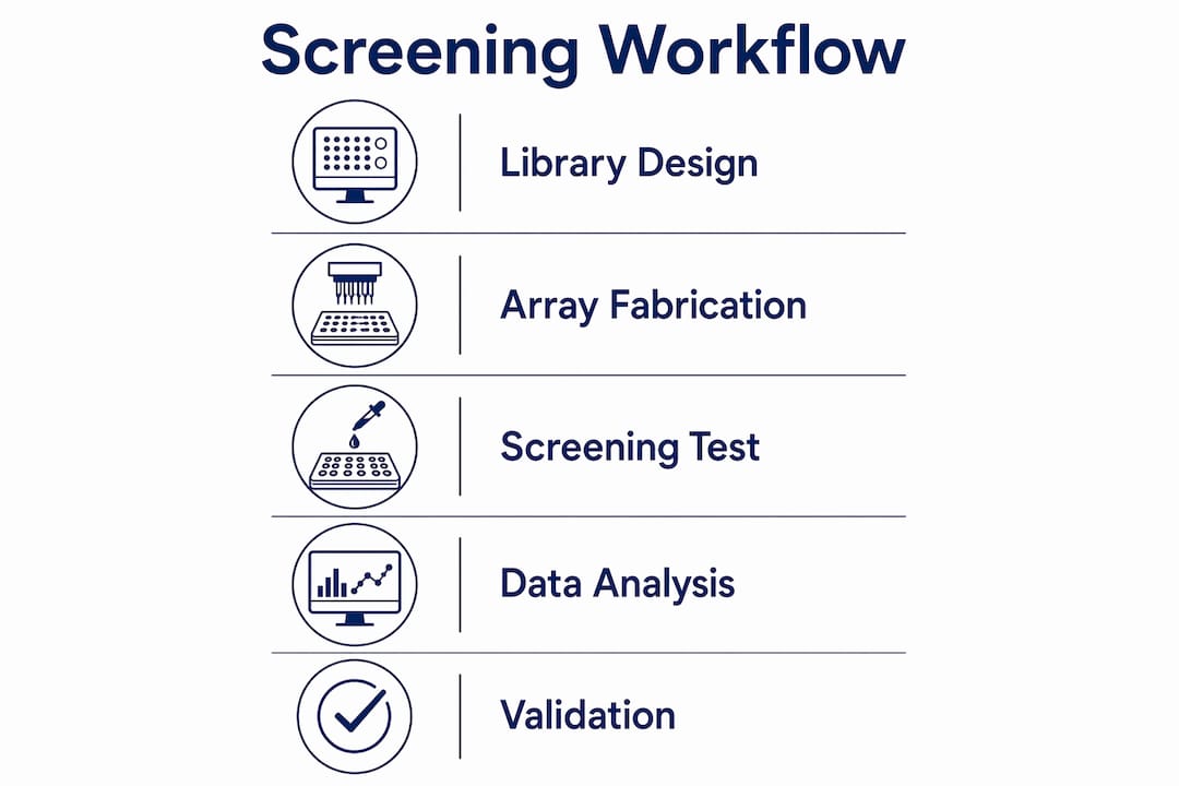
How peptide arrays work: design and fabrication for reliable screening
The physical construction of a peptide array determines the reliability of every data point it produces. Peptides are typically printed on NHS-ester functionalized glass slides using robotic microarray printers, and the choice of immobilization chemistry directly governs peptide orientation and binding stability. When orientation is poor, active binding sites face the slide surface rather than the solution phase, producing systematic false negatives that are difficult to distinguish from genuine non-binders.
Several fabrication variables require deliberate control before any screening campaign begins:
- Immobilization chemistry: Biotin-streptavidin coupling and PEG linkers preserve peptide accessibility and reduce nonspecific adsorption. NHS-ester chemistry offers covalent attachment but requires free amine groups positioned away from the binding epitope.
- Printing buffer composition: Buffer pH, ionic strength, and humidity during spotting affect peptide spreading, concentration uniformity, and surface density. Inconsistent printing conditions introduce spot-to-spot variability that inflates false positive rates.
- Tiling strategy: Overlapping peptide libraries, where each successive peptide shifts by one or a few amino acid residues, resolve epitope boundaries at single-residue resolution. The OmicsArray custom service from GeneCopoeia uses 1-amino-acid shift tiling to achieve this precision, printing up to approximately 11,288 unique peptides per slide.
- Multiplexing configuration: A single slide can accommodate up to 16 sample blocks, allowing multiple experimental conditions or patient samples to run in parallel. This multiplexing capacity reduces reagent consumption and inter-assay variability simultaneously.
- Post-print blocking: Saturating unreacted surface groups with ethanolamine or BSA after printing prevents nonspecific protein binding during the assay incubation step.
Pro Tip: When designing a tiling library for epitope mapping, include both N-terminally and C-terminally extended variants of your candidate sequence. This controls for linker proximity effects and confirms that the binding signal is sequence-specific rather than an artifact of surface attachment geometry.
Reproducibility across slides and print runs depends on batch-level quality controls analogous to those applied to synthetic peptides themselves. Researchers who treat array fabrication as a variable rather than a constant gain substantially more interpretable data from downstream fluorescence or mass spectrometry readouts.

What are the main applications of peptide arrays in screening?
Peptide array applications now span a broad range of research domains, each exploiting the platform’s capacity for parallel, quantitative measurement. The following applications represent the most established and productive uses in current biomedical and biotechnology research.
-
Oncology and tumor microenvironment profiling. Peptide microarrays enable parallel screening of thousands of sequences to identify motifs that modulate cancer cell adhesion and microenvironment interactions. This application is particularly valuable for identifying integrin-binding sequences and extracellular matrix mimetics that influence tumor progression.
-
High-throughput lead discovery in drug development. Fluorescence intensity on peptide microarrays serves as a quantitative proxy for peptide-target binding affinity, allowing researchers to rank hundreds of thousands of candidate sequences and prioritize leads for downstream synthesis and functional testing. This approach compresses what would otherwise require months of iterative synthesis into a single screening experiment.
-
Epitope mapping and antibody specificity profiling. A 2025 JoVE study demonstrates PTM-specific antibody testing on fluorophore-conjugated histone peptide arrays, distinguishing binding to target post-translational modifications from cross-reactive recognition of structurally similar marks. This level of specificity profiling is not achievable with standard ELISA formats.
-
Peptide biomarker detection in complex biological samples. Arrays incubated with serum or plasma can capture circulating antibodies or binding proteins against defined peptide sequences, enabling biomarker discovery without requiring prior knowledge of the analyte’s identity.
-
Integration with mass spectrometry and multi-omics platforms. User-defined peptide libraries paired with data-independent acquisition mass spectrometry (DIA-MS) recover greater than 75% of expected sequences for libraries exceeding 10,000 peptides, with detection sensitivity reaching low-femtomole concentrations in complex backgrounds. This positions peptide arrays as entry points into multi-omics workflows rather than standalone tools.
-
Post-translational modification (PTM) cross-reactivity mapping. Systematic testing of multiple PTM combinations reveals antibody cross-reactivity patterns that single-PTM testing cannot detect. This is directly relevant to researchers developing or validating epigenetic antibodies for chromatin immunoprecipitation or immunofluorescence applications.
The advantages of peptide arrays over phage display and computational screening are speed, quantitative output, and the ability to test chemically modified sequences including phosphopeptides, acetylated residues, and non-natural amino acids, which phage systems cannot accommodate.
What challenges affect interpretation of peptide array screening data?
Peptide array data requires careful interpretation because several technical and biological factors introduce ambiguity between a raw binding signal and a biologically meaningful interaction.
The most consequential source of error is immobilization chemistry. Orientation and immobilization chemistry can cause false negative or false positive results, and a 2026 review recommends confirmatory functional assays as a standard component of any array-based screening campaign. A peptide that binds strongly on an array may fail to engage its target in a cellular context because the array format constrains conformational flexibility or presents the sequence in an orientation that does not reflect the native protein surface.
The table below compares key factors that affect data quality and the corresponding mitigation strategies:
| Factor | Effect on data | Mitigation strategy |
|---|---|---|
| Poor peptide orientation | False negatives from blocked binding sites | Use flexible PEG linkers; test N- and C-terminal attachment |
| Nonspecific surface adsorption | False positives from background signal | Optimize blocking step; include scrambled peptide controls |
| Linear peptide format | Misses conformational epitopes | Validate hits with cyclic peptides or full-length protein assays |
| Insufficient tiling resolution | Epitope boundaries remain ambiguous | Apply 1-amino-acid shift tiling for precise mapping |
| Cross-reactive antibody binding | Overestimates specificity | Test full PTM combination matrix, not single modifications |
A second interpretive challenge is the linear peptide limitation. Arrays present sequences in isolation, without the tertiary structure context of the parent protein. Binding interactions that depend on a discontinuous epitope or a folded domain will not be captured, meaning array data describes linear sequence preferences rather than full protein binding landscapes. Researchers working on conformational epitopes must treat array hits as hypotheses requiring orthogonal validation methods such as cell-based functional assays, SPR with full-length proteins, or hydrogen-deuterium exchange mass spectrometry.
Pro Tip: Include at least three scrambled-sequence negative controls and one well-characterized positive control peptide in every array block. This establishes a reliable signal-to-noise baseline and allows you to calculate a statistically defensible hit threshold before analyzing experimental sequences.
Binding readouts from peptide arrays serve as hypothesis generators. Functional biological confirmation through orthogonal methods is the standard required for robust conclusions, not an optional follow-up step.
Practical strategies to optimize peptide array screening workflows
Designing an effective peptide array screening campaign requires decisions at multiple levels, from library composition to data analysis, each of which affects the quality and interpretability of results.
-
Define library scope before synthesis. Peptide length between 12 and 20 residues balances coverage of binding interfaces with synthesis yield and array density. For epitope mapping, overlapping 15-mers with a 3-residue shift provide good resolution without requiring the full single-residue tiling density that is more appropriate for precise boundary determination.
-
Match immobilization chemistry to your target. If your target protein binds the N-terminus of candidate peptides, C-terminal attachment to the slide surface is the correct choice. Mismatched chemistry systematically suppresses true hits. Consulting peptide chemistry resources before finalizing array design prevents this avoidable error.
-
Balance multiplex depth with peptide coverage. Multiplexing several samples per array reduces per-block peptide density. If your experiment requires 16 sample blocks, the available peptide slots per block drop proportionally, so library size must be adjusted accordingly. Prioritize the highest-value sequences when sample multiplex requirements are high.
-
Select detection methods aligned with throughput and sensitivity goals. Fluorescence imaging is the standard for initial screening due to speed and established quantification pipelines. Mass spectrometry integration, as demonstrated by the Pepyrus platform reported in Nature Biotechnology, extends sensitivity to low-femtomole detection and adds sequence-level confirmation to binding data.
-
Build a structured data analysis pipeline. Normalize fluorescence intensity values to positive controls within each block before comparing across slides. Apply a minimum signal-to-noise ratio threshold, typically 3:1 or higher, to define hits. Rank candidates by intensity and reproducibility across replicate spots before advancing to secondary assays.
-
Leverage commercial custom array services for complex projects. Platforms such as OmicsArray from GeneCopoeia offer PEPperPRINT laser printing with defined tiling options, PTM variants, and multiplexing configurations. For researchers without in-house printing infrastructure, these services provide access to high-throughput peptide screening capabilities without capital investment in robotic printing equipment.
Key takeaways
Peptide arrays generate reliable screening data only when immobilization chemistry, library design, and orthogonal validation are treated as integrated components of a single experimental system.
| Point | Details |
|---|---|
| Immobilization chemistry is critical | Linker choice and peptide orientation directly determine false positive and negative rates. |
| Tiling resolution defines epitope precision | Single-residue shift tiling resolves binding boundaries; coarser tiling only approximates them. |
| Fluorescence is a proxy, not a conclusion | Intensity data ranks candidates; functional assays confirm biological relevance. |
| Multiplexing imposes design trade-offs | More sample blocks per slide means fewer peptide slots per block; plan library size accordingly. |
| Mass spectrometry extends array utility | DIA-MS integration recovers over 75% of sequences in libraries exceeding 10,000 peptides at femtomole sensitivity. |
Why peptide arrays deserve earlier placement in discovery pipelines
From our perspective at Vertexpeptideslab, the most consistent observation across peptide array research is that these platforms are introduced too late in discovery workflows. Researchers frequently deploy arrays after candidate lists have already been narrowed by computational prediction or low-throughput binding assays, which means the platform’s primary advantage, screening thousands of sequences in parallel, is never fully realized.
We have also observed that the integration of peptide arrays with orthogonal assays is treated as a later-stage concern rather than a design requirement. Array data that is not paired from the outset with a plan for cell-based or SPR validation produces hits that stall at the confirmation stage. The experimental design should specify the validation pathway before the array is printed, not after the fluorescence data is in hand.
The convergence of AI-assisted library design and multi-omics data fusion is changing what peptide arrays can accomplish. Predictive models trained on binding interaction databases can now propose tiling libraries that maximize information density per slide, reducing the number of arrays needed to cover a given sequence space. Researchers who adopt this approach early in their programs will compress timelines considerably compared to those using static, manually designed libraries.
The practical advice we offer is direct: treat the array as the first quantitative filter in your pipeline, not a confirmatory tool. Design your library with the validation assay already specified, select your immobilization chemistry based on the target’s binding geometry, and build your controls into the array design itself. That sequence of decisions produces data worth acting on.
— Vertex
Explore Vertexpeptideslab’s research peptide resources

Vertexpeptideslab supports researchers conducting peptide array screening campaigns with laboratory-grade synthetic peptides verified by third-party testing and documented with full Certificates of Analysis confirming purity greater than 99%. Whether your workflow requires individual peptides for array validation, custom synthesis for tiling libraries, or reference compounds for positive controls, the Vertexpeptideslab research catalog provides batch-verified materials with full traceability documentation. Researchers can also consult our peptide database resources for sequence selection guidance and review our vendor evaluation criteria to assess supplier quality standards relevant to reproducible screening workflows. For laboratory research use only. Not for human or veterinary use.
FAQ
What is the role of peptide arrays in screening?
Peptide arrays enable parallel, high-throughput screening of thousands of peptide sequences against biological targets in a single experiment, generating quantitative binding, enzymatic, or specificity data. Their primary role is to identify candidate sequences for drug discovery, epitope mapping, and biomarker detection faster and at greater scale than sequential assay formats.
How does immobilization chemistry affect peptide array results?
Immobilization chemistry determines peptide orientation on the slide surface, and incorrect orientation blocks active binding sites, producing false negatives. Choosing linker chemistry based on the target’s known binding geometry, such as C-terminal attachment when the N-terminus is the functional region, is the primary control for this variable.
Can peptide arrays detect post-translational modifications?
Peptide arrays can distinguish antibody binding to specific PTMs from cross-reactive recognition of structurally similar modifications when the library includes systematic PTM combination variants. Testing a full matrix of PTM combinations, rather than a single modified sequence, reveals cross-reactivity patterns that single-peptide assays miss.
What detection methods are used in peptide microarray analysis?
Fluorescence imaging is the standard detection method for peptide microarray analysis due to throughput and established quantification pipelines. Mass spectrometry integration, particularly DIA-MS with user-defined peptide libraries, extends sensitivity to low-femtomole detection and adds sequence-level confirmation to binding data.
When should orthogonal validation follow peptide array screening?
Orthogonal validation should be planned before the array is printed, not after fluorescence data is collected. Array binding readouts are hypothesis generators; cell-based functional assays, SPR with full-length proteins, or additional biochemical methods are required to confirm that array hits represent biologically relevant interactions.